A reliable approach is to weigh out 100 g aliquots with a microbalance for use with individual experiments. Trichomonas vaginalis induces cytopathic effect on human lung alveolar basal carcinoma epithelial cell line A549. Publishing research using ab232855?
Rossitti HM, Dutta RK, Larsson C, Ghayee HK, Sderkvist P, Gimm O. Int J Mol Sci.

Crystal Violet Assay for Determining Viability of Cultured Cells. 2022 Jun 25;14(13):3125. doi: 10.3390/cancers14133125. An exception is M2 melanoma, which spreads rapidly on SN-peptide. Figure 1. K329-1000 is the same size as the 1000 test size of ab232855. Pull off remaining coating solution from each well with a yellow tip pipetter. Crystal Violet assay protocol summary:- remove culture medium- wash cells- add Crystal Violet staining solution- incubate for 20 mins- wash cells and air dry plate- add solubilization solution- analyze using a microplate reader at 570 nm absorbance. Agonists, activators, antagonists and inhibitors, Crystal violet, Cation-based violet dye (ab143095), MTT Assay Kit (Cell Proliferation) (ab211091), MTS Reagent, cell proliferation assay reagent (ab223881). Methods Mol Biol. A common working dilution for the laminin-1 positive control and BSA (Sigma A8412) negative control is 40 g/ml.

Activity decreases with storage, even frozen. Did you know that with a free Taylor & Francis Online account you can gain access to the following benefits?


I would say, if you normally grow your cells with serum, and you aren't trying to look at serum's influence on the cells, then you should grow them with serum as this is the normal condition for the cells. Add coating solution (100 l/well) with multipipetter to wells of a 96 well tissue culture plate (Costar #3595), cover, and place at 4C overnight.
violet crystal staining cell cells stained es colonies objective 40x feeder imaged growing When the calibrated method was employed to assess the attachment of Vibrio sp to polystyrene, stainless steel and copper, it gave a fairly reliable estimate of bacterial adhesion to these surfaces. Dissolve/dilute coating substrate in ddH2O at 4C.

Measure absorbance at 570 nm using a plate reader. Slowly add 100 l/well of serum-free medium down the side of each well. Remove non-adherent and loosely attached cells by either tapping the plate or gently washing the wells with PBS. The metabolism and mode of action of gentian violet. NOT FOR USE IN DIAGNOSTIC PROCEDURES" For licensing inquiries, please contact partnerships@abcam.com, Crystal violet Assay Kit (Cell viability). Chen L, Chiang YC, Chan LS, Chau WY, Lung ML, Kahn M, Lo KW, Mak NK, Lung HL. The remaining attached live cells are stained with Crystal violet. Invert plate and shake out coating solution.
programmed activates glioblastoma tumor temozolomide gbm knockdown proliferation adhesion FOIA Laminin value should be about 1.0 OD. Do you normally grow your cells with serum? *, *BSA background should be less than 0.1 OD. in a drawer. It depends- are you trying to look at the influence of serum on the cell attachment? There are currently no Customer reviews or Questions for ab232855.Please use the links above to contact us or submit feedback about this product. 2020 Oct 29;21(21):8072. doi: 10.3390/ijms21218072.
assays adhesion Assays are usually performed in triplicate or quadruplicate.

if i use serum-free media, is it also necessary to serum starve the cells? Thaw 10 x trypsin/EDTA (then dilute to 1 x in PBS), warm PBS and serum-free medium. After a wash step, the Crystal violet dye is solubilized and measured by absorbance at 570 nm. The site is secure. Please refer to protocols. Before my question is this: should i use serum-free media when i seed cells?
Pull off remaining wash from each well with pipetter. The amount of Crystal Violet staining in the assay is directly proportional to the cell biomass that is attached to the plate.
official website and that any information you provide is encrypted Invert plate gently onto an absorbent diaper pad. ab232855 has been referenced in 5 publications. Place in incubator for 30 - 60 min (37C).
colonization adherence 2009;522:203-10. doi: 10.1007/978-1-59745-413-1_14. With multipipetter, slowly add 100 l/well of freshly diluted 1% glutaraldehyde in PBS. With multipipetter, add 50 l/well of 0.5% Triton X-100 (diluted in ddH2O). is it alright to use the culture media (with serum) because to show how the cells adhere to the substrate? There was a highly significant positive linear relationship between crystal violet stained attached cells and the viable cell count of cells attached to aluminium panels (r = 0.9997; p < 0.001: n = 6). Store at -20C. If I test all of my conditions at RT under the same conditions, will this be a problem?Thanks.
bioengineering It is possible to investigate the nature of the molecules to which cells adhere in two different assays.
Pour cells in Reagent Reservoir (Costar # 4870), rock to suspend, remove 100 l/well with multipipetter and add to wells. Med Mycol. The method is relatively simple, sensitive, less time consuming and therefore many samples can be analysed in a short period of time. If incorrect, please enter your country/region into the box below, to view site information related to your country/region.
biofilm monocytogenes seca2 listeria mutant adhesion Novel Cell-Penetrating Peptides Derived From Scaffold-Attachment- Factor A Inhibits Cancer Cell Proliferation and Survival.

Dilute 7.5% BSA (Sigma A8412) to 1% in ddH2O. Assays were performed according to the kit protocol in triplicate. Repeat an additional time if required. The cell biomass can be used to infer levels of cell viability / cytotoxicity. Cell proliferation and invasion are regulated differently by EGFR and MRP1 in T-DM1-resistant breast cancer cells. Please contact us to place your order, or try again later. Get resources and offers direct to your inbox.

Age-related increase of kynurenine enhances miR29b-1-5p to decrease both CXCL12 signaling and the epigenetic enzyme Hdac3 in bone marrow stromal cells. Pull off remaining blocking solution from each well with a yellow tip pipetter. HelloI am doing dayly CV.
ecm assay adhesion mediated Read at OD 595.
transmembrane assay oulu 
Leave a blank well or wells for measuring background spreading on blocked plastic. *, *Cells in BSA negative control wells should be rare (if not, repeat wash). For the best experience on the Abcam website please upgrade to a modern browser such as Google Chrome. and transmitted securely. Access advice and support for any research roadblock, Full event breakdown with abstracts, speakers, registration and more, Explore the power of knock-out cell lines for your research, crystal-violet-assay-kit-cell-viability-ab232855.pdf. Fix cells in the wells to be used for determining 100% attachment value by addition of 100 L 5% (w/v) glutaraldehyde for 20 min at room temperature (or at 4C overnight if necessary). Examine plate in invert microscope. Add the diluted adhesion molecule to the wells of the microtiter plate (100 L/well).
Your browser does not have JavaScript enabled and some parts of this website will not work without it. Aspirate, add 200 L 10 mg/mL heat-denatured BSA in divalent cation-free PBS and incubate at room temperature for 30 min. An official website of the United States government.
adhesion suppresses silencing 1990;22(2-3):161-78. doi: 10.3109/03602539009041083. Register a free Taylor & Francis Online account today to boost your research and gain these benefits: The Journal of Bioadhesion and Biofilm Research, A simple method to assess bacterial attachment to surfaces, Marine Corrosion and Materials Research Division , National Institute of Oceanography , Dona Paula, Goa, 403004, India, /doi/pdf/10.1080/08927019509378289?needAccess=true. Dose-response curve of MCF7 (human breast adenocarcinoma cell line) cells to Doxorubicin for 72 hours determined by the Crystal violet Assay Kit (Cell viability) (ab232855).

In a second assay, an aliquot of cells is added to the well of a microtiter plate coated with a test adhesion molecule and the cells are allowed to attach.
The adhesion events can be divided into single cell and cell population analysis. 5 Howick Place | London | SW1P 1WG. We use cookies to improve your website experience. Invert plate gently onto an absorbent diaper pad.

While the blocking is underway, prepare a suspension of the cells to be examined. Sudjana AN, Carson CF, Carson KC, Riley TV, Hammer KA. People also read lists articles that other readers of this article have read. Invert plate gently onto an absorbent diaper pad. To learn about our use of cookies and how you can manage your cookie settings, please see our Cookie Policy. Dose-response curve of HepG2 (human liver hepatocellular carcinoma cell line) cells to Doxorubicin for 72 hours determined by the Crystal violet Assay Kit (Cell viability) (ab232855). National Library of Medicine All rights reserved. Cancers (Basel). In the first protocol presented in this unit, physical adhesion of a cell is assessed by determining the extent to which the cell spreads on a defined substrate--the plate is coated with the test substance, cells are added and allowed time to attach and spread, and the extent of spreading is assessed using phase contrast microscopy.
neuregulin dependent speeding regeneration fak facilitates schwann photomicrographs histogram erbb2 
2012 Nov;50(8):863-70. doi: 10.3109/13693786.2012.683540.
invasion cell assay assays ht 1080 determined cells were visualized staining invaded violet crystal 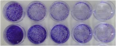
With multipipetter, slowly add 100 l/well of serum-free medium down the side of each well (tilt plate; PBS is not recommended for this wash).
violet assay crystal cell proliferation viability staining cells density mtt publication assays diagram government site. For the single cell study, the experiments are performed to analyze the interaction between the individual cell and the substrate, observing the morphological changes, studying the cellular migration, and measuring the traction forces. doi: 10.1101/pdb.prot087379.
lentivirus violet crystal ht1080 protocol ml cells ethanol Take a look at our BETA site and see what weve done so far.

Registered in England & Wales No. HHS Vulnerability Disclosure. Adherent cells in laminin-1 wells should be all spread. Allow to solubilize overnight at room temp. i guess i'm sort of confused as to how much serum masks the adhesion process (i've been under the assumption that it is needed to assist in adhesion).thanks!