11. Your heart is a strong, muscular organ situated slightly to the left of your chest. either the copyright owner or a person authorized to act on their behalf. When the SA node stimulates the heart, it initiated atrial systole. information contained in your Infringement Notice is accurate, and (c) under penalty of perjury, that you are

From the right atrium it is pumped to the right ventricle and then to the pulmonary arteries, which carry it to the lungs for reoxygenation.

The right atrium collects the impure blood from the vena cava and delivers it to the right ventricle. The valve which prevents the back flow of blood in the veins and lymph vessels is. Blood might flow back through the tricuspidvalve to the lungs through the right pulmonary artery. Because of this unique design and the assistance they receive from the heart they do not need valves to prevent backflow. This delivery is regulated by the tricuspid valve.
valves veins blood backflow vessels fig heart It prevents the backflow of blood to the left atrium when the left ventricle pumps blood through the aorta to the rest of the body. Superior vena cava: Receives blood from the upper body; delivers blood into the right atrium. Which of the followingwould happen if the chordae tendinae attached to the mitral valve were torn or damaged?

Because of this unique design and the assistance they receive from the heart. The tricuspid valve is on the right side of the heart. Meet Dr. Frank Bowen | Cardiothoracic Surgeon | Cooper University Health Care | Video Biography, Otolaryngology - Head and Neck Surgery (ENT), MitraClip Transcatheter Mitral Valve Repair. The mitral valve, after the left atrium, is where oxygenated blood arrives when it travels back to the heart from the left and right pulmonary veins. human cardiovascular system: Valves of the heart, To prevent backflow of blood, the heart is equipped with, https://www.britannica.com/science/valve-anatomy, University of Minnesota - Atlas of Human Cardiac Anatomy - Cardiac Valve Nomenclature. The aortic valve sits between the left ventricle and the aorta and prevents backflow of blood into the left ventricle after it contracts. Answer : a. The left ventricle collects the pure blood from the left atrium and delivers it to the aorta (main artery) from where it is pumped to the rest of the body. If you believe that content available by means of the Website (as defined in our Terms of Service) infringes one It is the pulmonary artery which carries deoxygenated blood to the lungs for oxidation from the right ventricle of heart.

People who have severe backflow may needvalve surgeryto prevent complications. In fact, most people who have MVP dont have backflow and never have any related symptoms or problems.

The mitral valve regulates the blood flow between the left atrium and the left ventricle. The left ventricle has a greater workload and is much more massive than the right ventricle but the two pump equal amounts of blood. The deoxygenated blood from the heart muscle is collected by the coronary veins and drained into the right atrium. Which of the following prevents the backflow of blood in venous circulation? What prevents backflow of blood inside the heart during contration ? If Varsity Tutors takes action in response to The pulmonary semilunar valve separates the right ventricle from the pulmonary artery and the aortic semilunar valve separates left ventricle from the aorta. Remember that "myo" refers to muscle and "cardio" refers to the heart. The most common types of arrhythmias are harmless. Certain conditions have been associated with MVP, including: Most people who have mitral valve prolapse (MVP) aren't affected by the condition. While every effort has been made to follow citation style rules, there may be some discrepancies. This delivery is regulated by the pulmonary valve. The bacteria attach to the valve and can cause a serious infection called infective endocarditis (IE). Right ventricle: Receives blood from the right atrium; pumps blood into the pulmonary artery.

University of Colorado Denver, Masters, Social Track your scores, create tests, and take your learning to the next level!

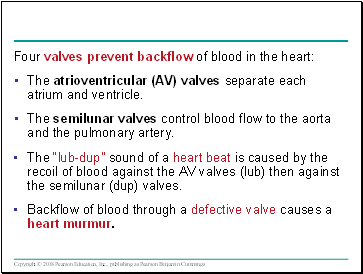
or more of your copyrights, please notify us by providing a written notice (Infringement Notice) containing The heart has two types of valves that keep the blood flowing in the correct direction. What does the heart provides your body with?
cardiovascular cram backflow ventricle valve, in anatomy, any of various membranous structures, especially in the heart, veins, and lymph ducts, that function to close temporarily a passage or orifice, permitting movement of a fluid in one direction only. The atrioventricular (AV) node, bundle of His, and Purkinje fibers are progressively lower in the conduction system and are not associated with P wave generation. That's more than 21 road trips between New York and Los Angeles! Recall that the right side of the heart deals with the oxygen-poor blood returned from the systemic circulation; this same blood is then pumped to the lungs to become oxygen-rich. your copyright is not authorized by law, or by the copyright owner or such owners agent; (b) that all of the The tricuspid valve is situated between the right atrium and right ventricle. Much of the time, MVP doesnt cause any problems.
 thoracic
 A description of the nature and exact location of the content that you claim to infringe your copyright, in \ Deoxygenated blood enters the right atrium through the superior and inferior vena cavae. Then, the tricuspid valve closes and the right ventricle contracts to pump the blood through the pulmonary valve into the pulmonary arteries, which carry oxygen-poorblood into the lungs to be oxygenated. Blood Flow through the Heart in 2 MINUTES. AV valves prevent backflow from the ventricles into the atria and semilunar valves prevent backflow from the aortic and pulmonary trunks into the ventricles.
The upper chamber is called the left atrium. These flaps normally help seal or open the valve. Fortunately.
backflow horizonal valve return non check backflow water brass female swing thread prevent dn50 unavailable pressure flash sorry player The sinoatrial node is the natural pacemaker of the heart. As a result, the atria aren't able to pump blood into the ventricles the way they should. Thus, if you are not sure content located The aortic semilunar valve prevents backflow from the aorta into the left ventricle. Gas exchange occurs between the alveoli and pulmonary capillaries. Mitral valve prolapse (MVP) is often detected during a routine physical exam whenyour doctorlistens to your heart with a stethoscope. The heart's four chambers pump in an organized manner with the help of electrical impulses that originate in the sinoatrial node (also called the "SA node").
Corrections?
backflow The adult heart weighs between 200 to 425 grams (7 to 15 ounces) and is about the size of your fist. 2007-2022 All Rights Reserved, ISEE Courses & Classes in San Francisco-Bay Area. Serious heart damage may occur when the coronary circulation is blocked. What prevents the backflow of blood in veins quizlet?
Depending on the severity of the mitral valve defect, mitral valve repair or mitral valve replacement may be needed. Laid end-to-end, your body's blood vessels would extend about 60,000 miles. COVID-19 Information:Testing |Vaccines|Vaccination for Camden Residents |VisitorPolicy. Our editors will review what youve submitted and determine whether to revise the article. Blood clots can occur because some blood "pools" in the atria instead of flowing into the ventricles.
valve mitral prolapse aortic ventricle left blood heart valves stenosis conditions known causes CVI most commonly occurs as the result of a blood clot in the deep veins of the legs a disease known as deep vein thrombosis (DVT).
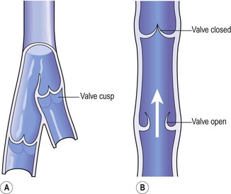
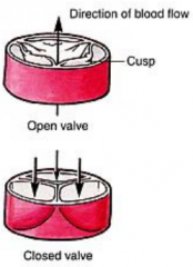
vein blood system valves cusps interior cardiovascular flow through valve direction figure basicmedicalkey One troublesome arrhythmia that MVP can cause isatrial fibrillation(AF). When veins lose integrity and the valves fail blood can collect and pool swelling the vessel. When it arrives in the left ventricle, it is pumped into the aorta to be delivered to the body. They tend to be mild but can worsen over time, mainly when complications occur. Other arrhythmias can be serious or even life threatening, such as ventricular arrhythmias. After passing through this valve, blood will be in the right ventricle. The mitral valve is between the left atrium and left ventricle. Stretched valve flaps can make a clicking sound as they shut. Its the muscle at the centre of your circulation system pumping blood around your body as your heart beats. Your Infringement Notice may be forwarded to the party that made the content available or to third parties such Please refer to the appropriate style manual or other sources if you have any questions. This delivery is regulated by the aortic valve. improve our educational resources. The valves between the atria and ventricles are called atrioventricular valves (also called cuspid valves) while those at the bases of the large vessels leaving the ventricles are called semilunar valves. 3. Cookies help us give you a better experience on our site. The left atrium collects the oxygenated blood from the lungs, via the pulmonary veins and delivers it to the left ventricle. Then, the ventricles contract to pump the blood out of the heart. Right atrium: Receives blood returning to the heart from the superior and inferior vena cava; transmits blood to the right ventricle, which pumps blood to the lungs for oxygenation. The heart acts a pump, delivering blood to the organs, tissues, and cells of your body through a complex network of arteries, arterioles, and capillaries. Inferior vena cava: Receives blood from the lower extremities, pelvis and abdomen, and delivers blood into the right atrium. 12. When backflow occurs, it can get worse over time and itcan change the hearts size and raise pressure in the left atrium and lungs. The blood travels through the body, and then returns to the vena cavae. pulmonary vein: One of four veins that carry oxygen-rich blood from the lungs to the heart. The sinoatrial node, SA node is called the heart's natural pacemaker that causes the atria to contract when the electrical impulse is released. Tricuspid valve: Allows blood to pass from the right atrium to the right ventricle; prevents blood from flowing back into the right atrium as the heart pumps (systole). The myocardium is the layer of the heart that contains the muscle cells. England Compared To Us State: What State Is England The Size Of? Medicines can treat troublesome MVP symptoms and help prevent complications. The mitral valve opens to allow the flow of blood from the left atrium into the left ventricle, the left atrium contracts to help.

"Epi" means above or over, "endo" means within or inner; the epicardium and endocardium are serous membranes that comprise part of the pericardium, which protects and lubricates the heart. Some people's valves are abnormal in more than one way. Lack of blood flow can damage the brain, heart, and other organs. Backflow also increases the risk ofinfective endocarditis(IE). The flaps of the mitral and tricuspid valves open to let blood through. Blood flows from your right atrium into your right ventricle through the. which specific portion of the question an image, a link, the text, etc your complaint refers to; What structures prevent backflow of blood into the heart quizlet? There is a specialized group of cardiac cells responsible for initiating this action potential throughout the heart. With each heartbeat, the atria contract and push blood into the ventricles. An identification of the copyright claimed to have been infringed; The mitral valve separates the left atrium from the left ventricle, and is also known as the bicuspid valve. Even people who do have symptoms may not need treatment. Mitral valve prolapse (MVP) is a condition in which the hearts mitral valve doesnt work well. Mitral valve: Allows blood to flow into the left ventricle; prevents blood from flowing back into the left atrium. Both the right and left sides of the heart must pump the same volumes since all blood from the right side returns to the left side after passing through the lungs. The aortic valve is between the left ventricle and the aorta.

They are involved in signal mediation and ventricular systole, which corresponds with the QRS complex. Diuretics (fluidpills) to remove excess sodium and fluid in your body and lungs. To understand the anatomy and function of the heart, we have divided the heart into two sections - Exterior and Interior. The cardiac sphincter divides the esophagus from the stomach, and is actually part of the digestive system. Aortic valve: Allows blood to pass from the left ventricle to the aorta; prevents backflow of blood into the left ventricle. In turn, veins bring nutrient-depleted blood back to the heart. On average, your heart will beat 100,000 times and pump about 2,000 gallons of blood each day. When MVP does cause signs and symptoms, they may include: MVP symptoms can vary from one person to another. The heart has a total of four chambers: right atrium, right ventricle, left atrium and left ventricle. If you've found an issue with this question, please let us know.
 veins valves blood venous valve prevent 3b ensure flow
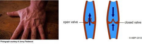
 How do the dimensions of health interact with each other? 9. Valves also help blood travel back to the heart against the force of gravity. The oxygen-rich blood from the lungs comes to the left side of the heart, where it will be pumped to the body tissues. pulmonary artery: A blood vessel that carries blood from the heart to the lungs where the blood picks up oxygen and then returns to the heart.

6.
mediastinum anatomy gross pulmonary lecture prevents ventricle blood heart into aorta left flashcards cram artery right Each side has an atrium (which receives blood as it enters) and a ventricle (from which blood is pumped out). 5. Medicines such as flecainide and procainamide to regulate your heart rhythms. University of Rochester Medical Center - Health Encyclopedia - What are Heart Valves. In order for the entire heart to contract in unison, there needs to be a conduction pathway that sends an action potential throughout the entire heart muscle at once. As part of the pulmonary circulation, the pulmonary artery carries the de-oxygenated blood from the right ventricle to the lungs for oxygenation. Your heart is a muscle, which unlike other muscles of the body has to work around the clock to keep your blood circulating. Which blood vessel carries blood for oxidation? This delivery is regulated by the mitral valve.
valve aortic semilunar function blood valves heart semi into lunar pulmonary body backflow definition study circulatory prevent aorta arteries located